Introduction
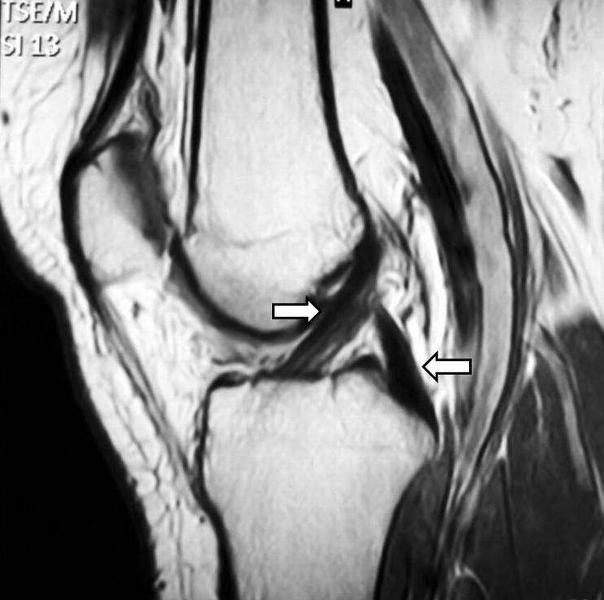
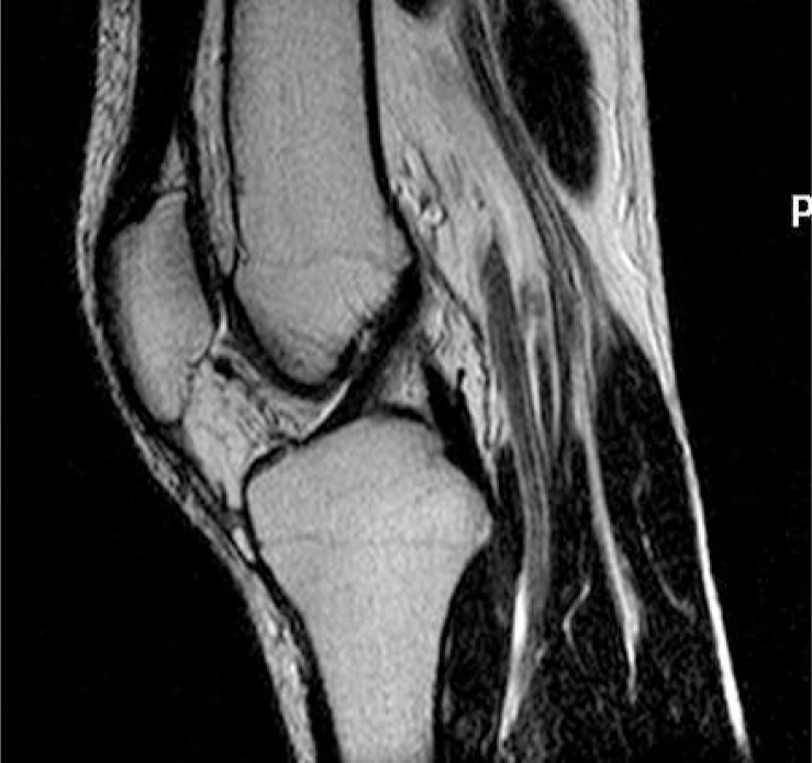
The anterior cruciate ligament (ACL) is vital in maintaining knee joint stability, especially in preventing anterior tibial translation and rotation.1 It is one of the two cruciate ligaments in the knee, the other being the posterior cruciate ligament (PCL) (see Figures 1 and 2).2,3 They are named "cruciate" due to the cross-like formation these ligaments create.4 The terms "anterior" and "posterior" refer to their attachment points on the tibia.4
Embryology
The ACL begins its development around the sixth week of gestation, even before joint cavitation occurs.1 Due to its embryological development, the ACL remains extrasynovial throughout its course while being intra-articular, due to a surrounding fold of synovium which originates from the posterior of the joint.1
Anatomy
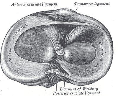
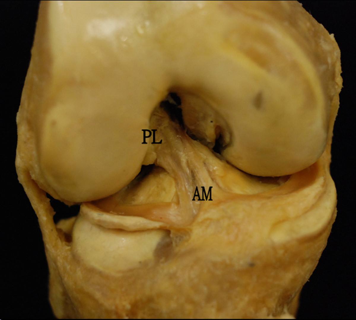
Both the ACL and the PCL are located within the intercondylar region of the knee joint.1 The ACL arises from the medial part of the anterior intercondylar area of the tibia.4,5,7 The ACL sends fibres anteriorly beneath the anterior (transverse) intermeniscal ligament, with some blending with the anterior and posterior horns of the lateral meniscus.1 From its tibial attachment, the ACL courses upwards, laterally, and posteriorly to attach to the posterior aspect of the medial side of the lateral femoral condyle.4,7 The tibial attachment of the ACL is stronger than its femoral attachment, although, the PCL is the stonger of the two cruciate ligaments overal.4,5 The ACL is comprised of two distinct bundles observable on MRI: a smaller anteromedial bundle (AMB) and a larger posterolateral bundle (PLB).1,5,6 These two bundles are surrounded by connective tissue, seen as high-signal on MRI making the distinction between the bundles clear, while also having separate insertion sites.1,6-8
The AMB arises from the most anterior and proximal aspect of the femoral attachment, while the PLB originates from the posterodistal aspect of the femoral attachment.1 At the tibial attachment, the AMB inserts anteromedially, and the PLB inserts posterolaterally.1
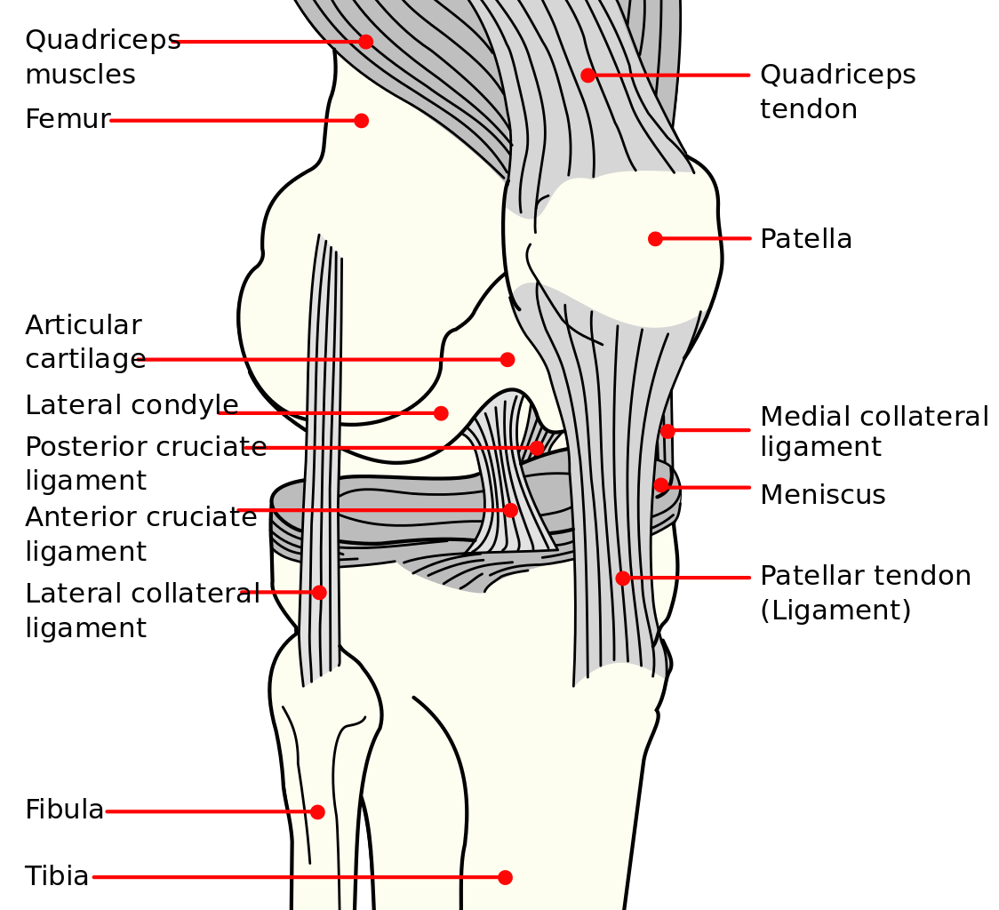
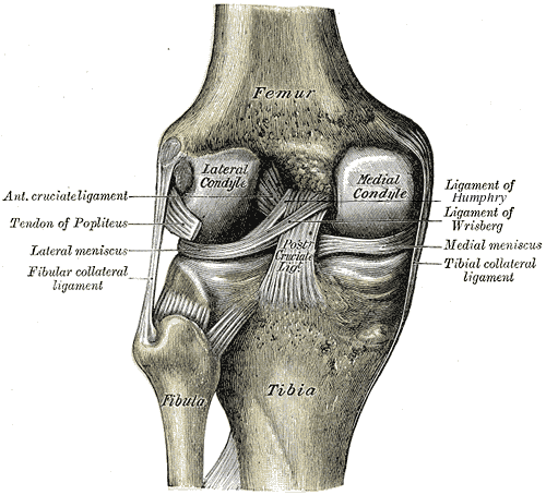
Refer to Figures 3-6 for images of the knee ligaments and menisci, as well as the bundles of the ACL.9-12
Blood Supply and Innervation
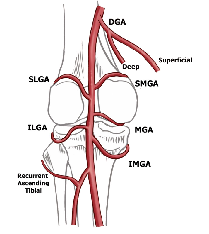
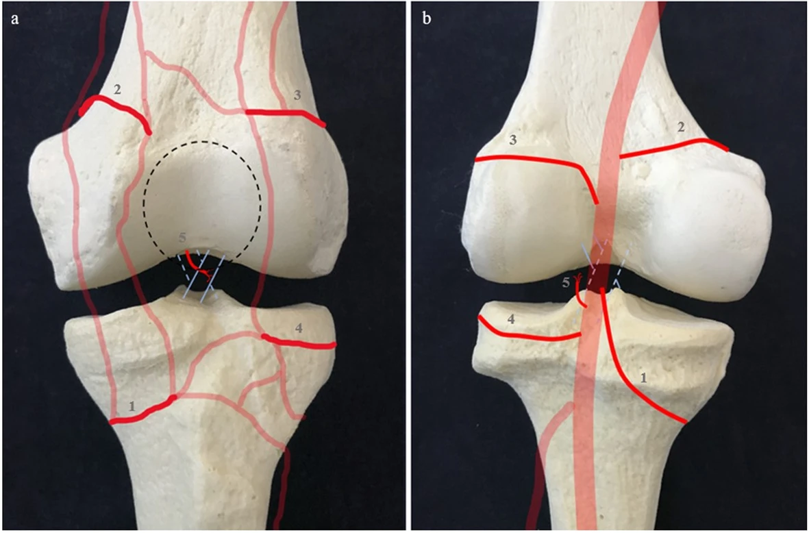
Blood Supply
Innervation
The ACL receives fibres from the posterior articular branches of the tibial nerve.1
Variations
Some studies suggest that the ACL is composed of three bundles: AMB, PLB, and an intermediate bundle.1,8 Others indicate that in up to 26% of individuals, the ACL can be a single bundle.8
Imaging
The ACL differs from other ligaments by appearing heterogeneous in signal on MRI. This characteristic is beneficial in visualising and diagnosing ACL injuries.7
Clinical Relevance
ACL Tears:
References
- 1. Duthon VB, Barea C, Abrassart S, Fasel JH, Fritschy D, Me´ne´trey J. Anatomy of the anterior cruciate ligament. Knee Surg Sports Traumatol Arthrosc. 2006;14(3):204-213. doi:10.1007/s00167-005-0679-9.
- 2. Lykissas MG, Mataliotakis GI, Paschos N, Panovrakos C, Beris AE, Papageorgiou CD. MRT ACL PCL 01. Wikimedia Commons. Available at: https://commons.wikimedia.org/wiki/File:MRT_ACL_PCL_01.jpg. Accessed August 10, 2024. Licensed under CC BY-SA 2.0.
- 3. Nenezic D, Kocijancic I. The value of the sagittal-oblique MRI technique for injuries of the anterior cruciate ligament in the knee. Radiol Oncol. 2013;47(1):19–25. doi:10.2478/raon-2013-0006. PMCID: PMC3573830.
- 4. Gray H. Anatomy of the Human Body. 21st ed. Revised and edited by Lewis WM, MD. Philadelphia, PA: Lea & Febiger; 1918.
- 5. Ma Y, Mandell J. Chapter 13: Musculoskeletal Imaging. In: Robinson-Weiss C, Malone FE, Sun EX, Shi J, Jhala K, Matalon SA, eds. Core Radiology: A Visual Approach to Diagnostic Imaging. 2nd ed. Cambridge: Cambridge University Press; 2021:908-1083. Volume 1 ISBN 978-1-108-98444-7; Volume 2 ISBN 978-1-108-98445-4. doi:978-1-108-96645-0.
- 6. Siegel L, Vandenakker-Albanese C, Siegel D. Anterior Cruciate Ligament Injuries: Anatomy, Physiology, Biomechanics, and Management. Clin J Sport Med. 2012;22(4):349-355. doi:10.1097/JSM.0b013e3182580cd0.
- 7. Ansede G, Mitchell AWM, Healy JC. Chapter 16: The Lower Limb. In: Butler P, Mitchell A, Healy J, eds. Applied Radiological Anatomy. 2nd ed. Cambridge University Press; 2012:319-365. ISBN 978-0-521-76666-1.
- 8. Morales-Avalos R, Torres-González EM, Padilla-Medina JR, Monllau JC. ACL anatomy: Is there still something to learn? Rev Esp Cir Ortop Traumatol. 2024;68(4):422-427. doi:10.1016/j.recot.2023.02.005. PMID: 36787832.
- 9. NicDumZ. Diagramme genou couleur. Wikimedia Commons. Available at: https://commons.wikimedia.org/wiki/File:Diagramme_genou_couleur.png. Accessed August 10, 2024. Licensed under CC BY-SA 3.0.
- 10. Carter HV. Gray's Anatomy - Gray348-2. Wikimedia Commons. Available at: https://commons.wikimedia.org/wiki/File:Gray348-2.png. Accessed August 10, 2024. Public domain.
- 11. Carter HV. Gray's Anatomy - Gray349. Wikimedia Commons. Available at: https://commons.wikimedia.org/wiki/File:Gray349.png. Accessed August 10, 2024. Public domain.
- 12. Lam MH, Fong DT, Yung PS, Ho EP, Chan WY, Chan KM. ACLI 18. Wikimedia Commons. Available at: https://commons.wikimedia.org/wiki/File:ACLI_18.jpg. Accessed August 10, 2024. Licensed under CC BY-SA 2.0.
- 13. Callese T, Cusumano LR, Redwood KD, et al. Classification of genicular artery anatomic variants using intraoperative cone-beam computed tomography. CardioVascular and Interventional Radiology. March 2023;46(5). Available at: https://doi.org/10.1007/s00270-023-03411-3. Accessed August 10, 2024. Licensed under CC BY 4.0.
- 14. Sinno E, Cavallo AU, Cera G, et al. Magnetic resonance imaging landmarks for preoperative localization of inferior medial genicular artery: a proof of concept analysis. J Exp Orthop. 2020;7(73). Available at: https://doi.org/10.1186/s40634-020-00288-w. Accessed August 10, 2024. Licensed under CC BY 4.0.